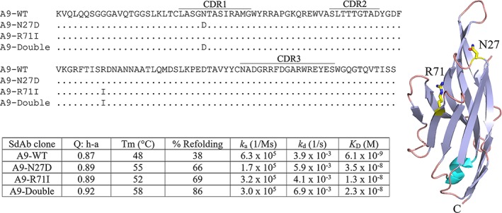
Figure 1.

Sequences of A9 (PDB 4idl) and the mutants used in this study are shown. Changes in the mutants are indicated. The CDR identifications follow the usage in Bekker et al.4 The crystal structure of A9 is shown with the mutated amino acids rendered as a stick model. The table shows the melting point and affinity data reported in this study. “Q:h‐a” is the Q score for the set “hydrophilic ‐all” and is taken from Bekker et al.4
