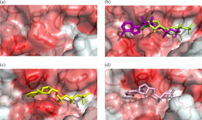
Figure 6.

Opening of the hydrophobic pocket at the InvCΔ79 ATP‐binding site and comparison with T3SS ATPase orthologues. Surface and ribbon representations colored by higher (red) to lower (white) hydrophobicity of amino acids showing (a) the Salmonella InvCΔ79 apo‐form, (b) InvCΔ79 in presence of ADP (purple) and ATPγS (lime), (c) the Shigella Spa47Δ83 with ATPγS (PDB ID: 5ZT1) and (d) the E. coli EscNΔ102 with ADP (PDB ID: 2OBM). Ligands and side chains of amino acids stabilizing the ligands are depicted as sticks in the same orientation as in Figure 5
