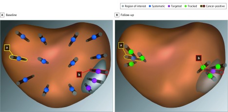
Figure 1. Systematic, Targeted, and Tracked Prostate Biopsy Schematic.
Biopsy approaches shown on reconstructed images from magnetic resonance imaging–ultrasonography fusion device. A, Region of interest with 3 targeted biopsy cores, 1 containing cancer tissue (b). Systematic biopsy with 1 biopsy core containing cancer tissue (a). B, Follow-up with tracked biopsy cores from the cancerous site within (b) and distant from (a) the region of interest. Tracked biopsies were placed to circumscribe a previously abnormal site.

