Fig. 6.
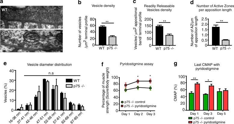
Reduced presynaptic vesicles in p75NTR−/− mice contribute to impaired synaptic transmission and muscle strength. a–e Diaphragm NMJs from 2 to 4 months old WT and p75NTR−/− (n = 3) mice were analyzed by transmission electron microscopy. a Representative images of WT and p75NTR−/− NMJs. Scale bar: 500 nm. b The total number of vesicles in each axon terminal was quantified as a fraction of the synaptic terminal area. c To quantify the RRP of vesicles, the number of vesicles within a 480 nm strip directly across active zones was counted. d Active zones (AZ) were quantified as electro dense regions located in direct apposition to postsynaptic secondary folds. The results are expressed as a function of the length of pre/postsynaptic apposition. e Vesicle diameter was measured in 40–110 vesicles per nerve terminal (n = 3–4 NMJs per mice). f–g Pharmacological inhibition of acetylcholinesterase with daily subcutaneous administration of pyridostigmine bromide partially rescued muscle strength (n = 5) (f) and NMJ transmission (n = 4) (g) of p75NTR−/− mice. The results represent the mean ± SEM. n.s., non-significant, *p < 0.5; **p < 0.01, unpaired t-test. b–d, f, g, or two-way ANOVA (e)

