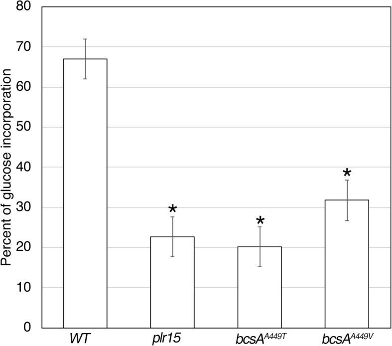
Fig. 5.

Comparison of radiolabeled glucose incorporation into the cellulose fraction of pellicles from K. xylinus wild type and plr mutants. Measurements were determined after bacterial cultures were incubated in 14C-glucose for five hours. The vertical axis is expressed as percentage of 14C-glucose detected in the insoluble fractions divided by the soluble plus insoluble fractions. Asterisks indicate statistically significant differences compared to wild type according to two-tailed student t-test (p < 0.05). Values are the mean ± standard deviation (n = 6)
