
Fig. 4.
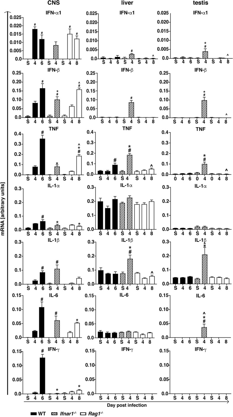
Expression of pro-inflammatory cytokine genes in the CNS, liver, and testis of WT, Ifnar1−/−, and Rag1−/− mice. Densitometric quantifications of RPAs for IFN-α1, IFN-β, TNF, IL-1α, IL-1β, IL-6, and IFN-γ mRNA. Cytokine mRNA levels were normalized to the housekeeping gene L32. Values are shown as mean ± SEM. #p < 0.05 when compared with sham-injected mice, *p < 0.05 when compared with day 6 (peak disease) in WT mice, ^p < 0.05 when compared with day 4 (peak disease) in Ifnar1−/− mice, as determined by one-way ANOVA