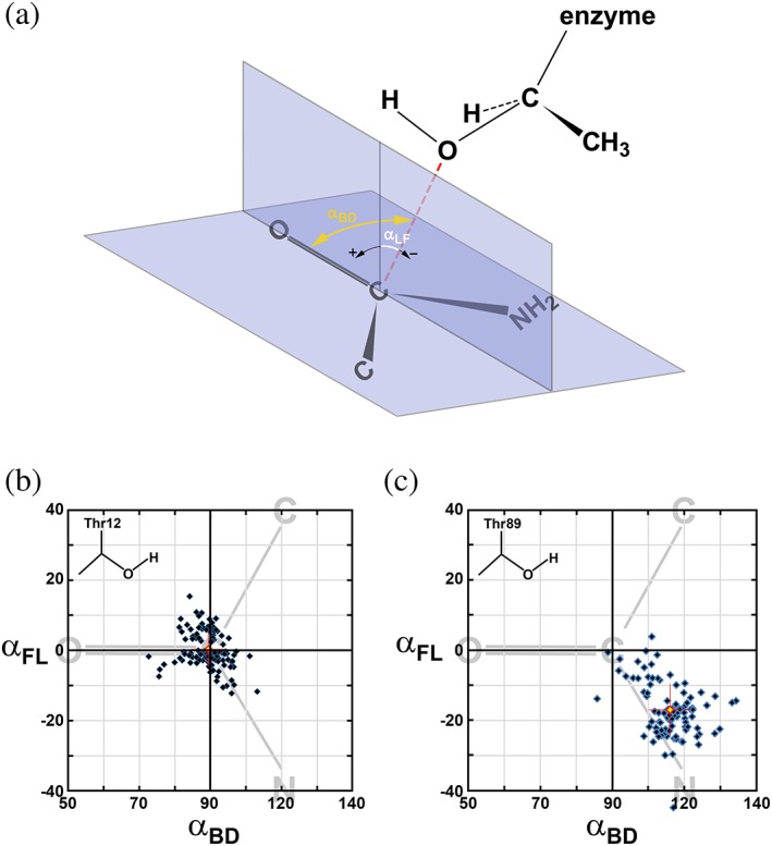
Figure 2.

Distribution of putative Bürgi–Dunitz (αBD) and Flippin–Lodge (αFL) angles for the Thr12(Oγ) and Thr89(Oγ) atoms in l‐asparaginases. (a) Angle between the direction of nucleophilic attack (red dashed line) and the carbonyl bond corresponds to αBD. Angle between the direction of nucleophilic attack and its projection on the plane perpendicular the carboxyamide group is defined as αFL. (b) Distribution αBD and αFL for 111 independent active sites of l‐asparaginases complexes with a substrate and calculated based on assumption that Thr12(Oγ) atom is a nucleophile. Each blue diamond represents a single active site and the red diamond represents the average (αBD, αFL). The latter is accompanied by red crosshairs representing values of standard deviations for both angles. The area of distribution is limited to the ranges of 50–140° for αBD and −40 to 40° for αFL which encompasses all data points. This area is coplanar with the carboxyamide group that is schematically represented by simplified formula (shown in gray) and oriented appropriately relative to αBD and αFL axes. (c) The same distribution as described in Panel B, but representing αBD and αFL for 95 independent active sites of l‐asparaginases complexes with a substrate and calculated based on an assumption that Thr89(Oγ) is the nucleophile
