
Figure 2.
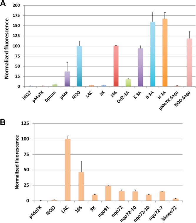
Characterization of different promoters in the pMoT system. (A) Fluorescence levels measured for the indicated promoters transformed into Tth. HB27, Tth HB27 strain with no plasmid; pMoTK, plasmid without promoter and sIFP gene; Δprom, construct without promoter; pMK, pMKnqosGFP; NQO, pMoTK with Pnqo driving sIFP; LAC, pMoTK with Lac promoter driving sIFP; 3K, idem with 3K promoter; 16S, idem with rRNA 16S promoter; Ori2-3A, pMoTK with nqo 3A promoter and pTT8 origin of replication; K 3A, pMoTK with nqo 3A promoter; B 3A, pMoTB with nqo 3A promoter; H 3A, pMoTH with nqo 3A promoter; C + Δago, pMoTK with Pnqo-sIFP transformed in Δago strain; C-Δago, pMoTK transformed in Δago strain. (B) Fluorescence levels measured for the indicated promoters in E. coli cultured at 37 °C. nqo variants as indicated in Figure 3B.