
Figure 2.
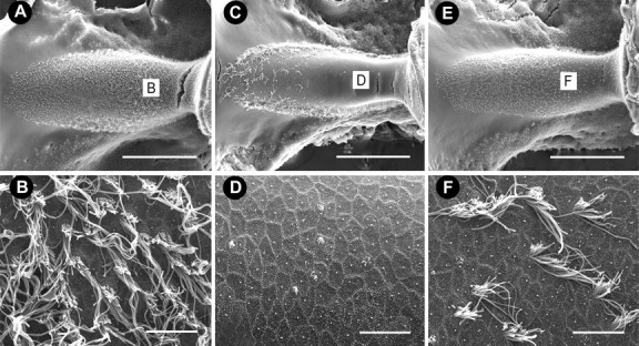
Scanning electron micrographs of the horizontal crista ampullaris. A, Low magnification of the crista from an untreated animal with numerous hair cells throughout the sensory epithelium. B, High magnification of the central apical region. C, Low-magnification image from an animal treated with 5 d of streptomycin (1200 mg/kg) showed that surviving hair cells were present only at the edge of the epithelium. Hair cells with mature stereocilia are absent from the central apical region of the crista (C, D). E, Low-magnification image from an animal treated with 5 d of zVAD (1.5 mg/kg) and streptomycin. F, Central apical region of horizontal crista ampullaris shows only moderate hair cell loss. Scale bars: low magnification, 500 μm; high magnification, 10 μm.