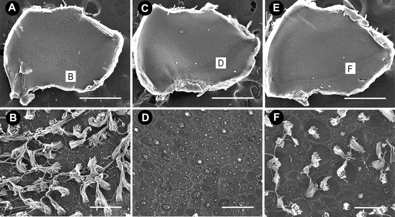
Figure 3.

Scanning electron micrographs of utricles. A, Low-magnification image of a control utricle with densely populated hair cells. B, High-magnification image of the striolar region of the same utricle as shown in A. C, After 5 d of streptomycin treatment, hair cell damage is predominately localized to the striolar region with extensive stereociliary loss evident along the entire length of the striola (D). E, After 5 d of zVAD and streptomycin treatment, some hair cell loss is evident in the striola, but most hair cells are still present. Scale bars: low magnification, 500 μm; high magnification, 10 μm.
