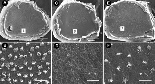
Figure 4.

Scanning electron micrographs of saccules. A, Low-magnification image of a control saccule, densely populated by hair cells. B, High-magnification image of the striolar region of the same saccule as shown in A. C, After 5 d of streptomycin treatment, hair cell damage is predominately localized to the striolar region with extensive stereociliary loss evident along the entire length of the striola (D). E, After 5 d of zVAD and streptomycin treatment, hair cell loss is evident in the striolar region (F), but many hair cells are still present when compared with animals treated with streptomycin alone. Scale bars: low magnification, 500 μm; high magnification, 10 μm.
