
Figure 1.
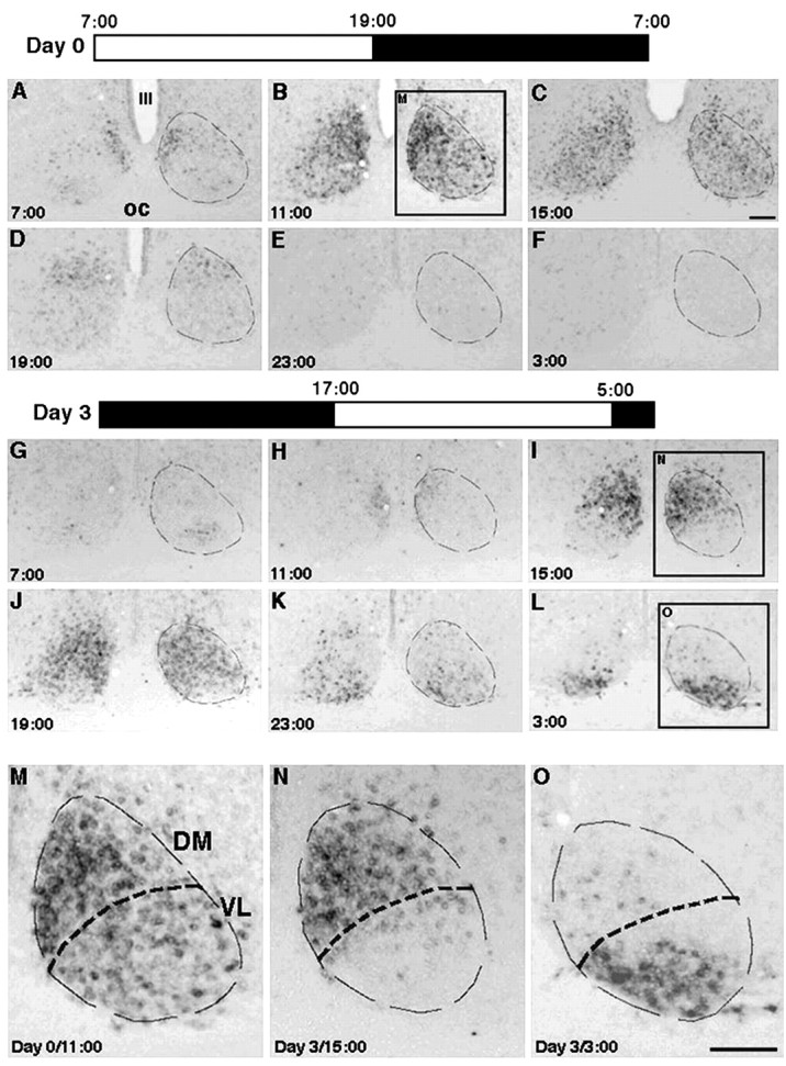
rPer1 expression in the SCN before and after a shift in the light/dark cycle. Coronal sections of the rat bilateral SCN were processed for in situ hybridization using a rPer1 probe. Dashed lines indicate the SCN boundaries and denote DM and VL subdivisions within the SCN. Scale bar, 200 μm. A–F, M, Brains from rats maintained on the light/dark cycle shown as Day 0. Rat brains were collected every 4 hr over a 24 hr period. Clock time is denoted in each panel. Before the photoperiod shift, the subdivisions of the SCN are synchronized with respect to their rPer1 expression. This is particularly evident when viewed at a higher magnification (M), at which the DM and VL SCN are easily delineated. G–L, N, O, Brains taken every 4 hr from rats on the third day after a 10 hr shift in the light/dark cycle. The new photoperiod is shown in Day 3. The abrupt shift in the lighting cycle causes the SCN subregions to become out of phase with each other. This is especially clear at higher magnification (N, O). OC, Optic chiasm; III, the third ventricle.