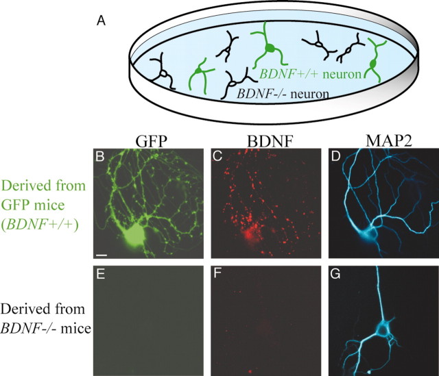
Figure 1.
Chimera cell culture prepared from two types of transgenic mice. A, Schematic illustration of chimera culture of cortical neurons derived from BDNF-/- and GFP mice. The latter neurons were labeled with GFP and had the potential to express endogenous BDNF (BDNF+/+). B, GFP image of a cortical neuron derived from a GFP mouse. C, Immunocytochemical BDNF image of the neuron shown in B. D, MAP2 image of the neuron shown in B and C. E, No GFP fluorescence signal in a cortical neuron derived from a BDNF-/- mouse was observed. This neuron was located in the same dish as above. F, No BDNF immunoreactivity in the neuron shown in E was observed. G, MAP2 image of the neuron shown in E and F. Scale bar: (in B) B–G, 10 μm.

