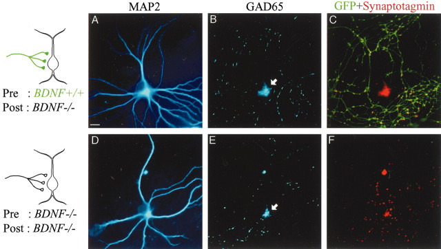
Figure 5.
Promoted growth of dendrites of GABAergic neurons by presynaptic BDNF. A, MAP2 image of a neuron contacted by a GFP-positive axon (BDNF+/+), as schematically shown on the left. B, GAD65 image of the same frame as in A. The arrow shows the cell body. C, Superposition of GFP images (green) and synaptotagmin images (red) of the same frame as A and B. D, MAP2 image of a neuron contacted by a GFP-negative axon (BDNF-/-), as schematically shown on the left. E, GAD65 image of the same frame as in D. The arrow shows the cell body. F, Superposition of GFP images (green) and synaptotagmin images (red) of the same frame as D and E. Because both the presynaptic and postsynaptic neurons were GFP negative, there was no green signal. Scale bar: (in A), 10 μm.

