
Figure 3.
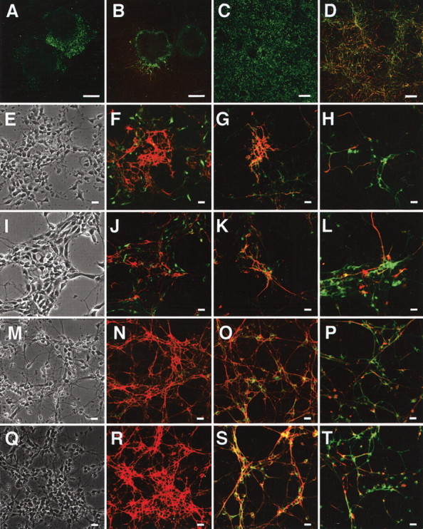
Analysis of morphology and marker expression in ES-derived cells. A, B, Day 7 of wild-type EBs attached on fibronectin-coated dishes and stained with anti-nestin antibody (A, green) or both anti-MAP2 (green) and anti-NF-M (red) antibodies (B). C,D, Day 9 of cells derived from wtES cells cultured on fibronectin-coated dishes and stained with anti-nestin antibody (C, green) or both anti-MAP2 (green) and anti-NF-M (red) antibodies (D). E–H, Day 11 (postpolyO/L day 2) of wtES-derived cells stained with anti-nestin (green) plus anti-E-NCAM (red) antibodies (F), anti-MAP2 (green) plusanti-E-NCAM antibodies (red) (G), or anti-MAP2 (green) plus anti-NF-M (red) antibodies (H). E is a phase-contrast image of F. Representative fields are indicated. I–L, Day 11 of knock-in ES-derived cells stained with anti-nestin (green) plus anti-E-NCAM (red) antibodies (J), anti-MAP2 (green) plus anti-E-NCAM (red) antibodies (K), or anti-MAP2 (green) plus anti-NF-M (red) antibodies (L). I is a phase-contrast image of J. M–P, Day 13 (post-polyO/L day 4) of wtES-derived cells stained with anti-nestin (green) plus anti-E-NCAM (red) antibodies (N), anti-MAP2 (green) plus anti-E-NCAM (red) antibodies (O), or anti-MAP2 (green) plus anti-NF-M (red) antibodies (P). M is a phase-contrast image of N. Q–T, Day 13 of knock-in ES-derived cells stained with anti-nestin (green) plus anti-E-NCAM (red) antibodies (R), anti-MAP2 (green) plus anti-E-NCAM (red) antibodies (S), or anti-MAP2 (green) plus anti-NF-M (red) antibodies (T). Q is a phase-contrast image of R. Scale bars: A, B, 200 μm; C, D, 100 μm; E–T, 25 μm.