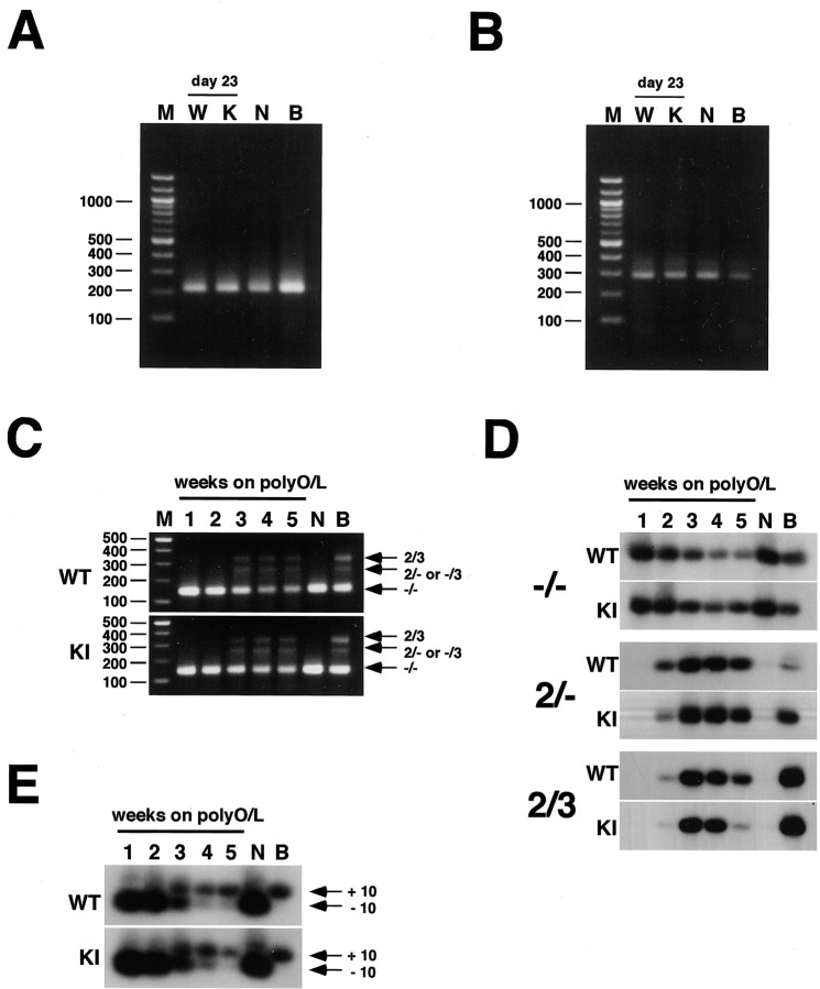
Figure 6.
Expression of tau isoforms. A, RT-PCR analysis of the splicing at exons 6 and 8 using the primer set 1 in neurons derived from wild-type or knock-in ES cells after 23 d of differentiation. B, RT-PCR analysis of the splicing at exons 4a, 6, and 8 using the primer set 2 in neurons derived from wild-type or knock-in ES cells after 23 d of differentiation. C, RT-PCR analysis of the splicing at exons 2 and 3 in neurons derived from wild-type (WT) or knock-in (KI) ES cells during the period from 1 to 5 weeks after plating on polyO/L dishes. -/-,2/- or -/3, and 2/3 represent tau isoforms lacking both exons 2 and 3, containing either exon 2 or exon 3, and containing both exons 2 and 3, respectively. D, RT-PCR analysis of the splicing at exons 2 and 3 in neurons derived from wild-type (WT) or knock-in (KI) ES cells during the period from 1 to 5 weeks after plating on polyO/L dishes. -/-,2/-, and 2/3 represent tau isoforms lacking both exons 2 and 3, containing exon 2 but not exon 3, and containing both exons 2 and 3, respectively. PCR products were submitted to Southern blot analysis using 301 bp fragment of tau. E, RT-PCR analysis of the splicing at exon 10 in neurons derived from wild-type (WT) or knock-in (KI) ES cells during the period from 1 to 5 weeks after plating on polyO/L dishes. +10 and -10, tau isoforms containing and lacking exon 10, respectively. PCR products were submitted to Southern blot analysis using 164 bp fragment of tau. Each of the results was reproduced at least three times.

