
Figure 4.
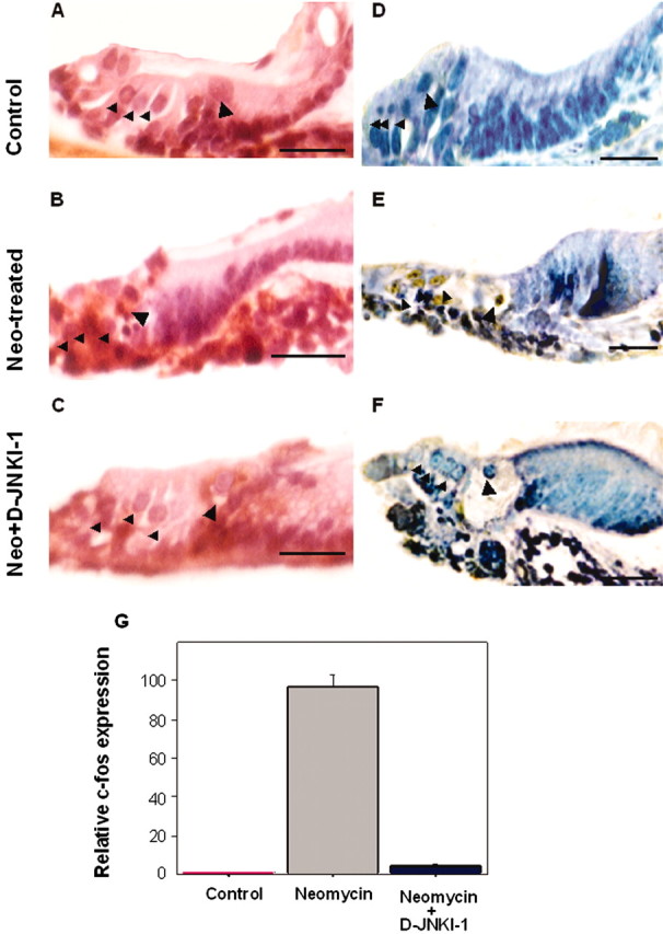
Neomycin exposure caused both hair cell loss and c-Jun phosphorylation in sensory cell nuclei in organ of Corti explants. Transverse plane tissue sections of P3 mouse organ of Corti explants. A, D, Untreated, control explants. B, E, Explants exposed to 1 mm neomycin for 24 hr. C, F, Explants exposed to 1 mm neomycin in the presence of D-JNKI-1 (2 μm) for 24 hr. A–C, Sections stained with hematoxylin and eosin. Nuclear condensations were present in some of the remaining hair cells in the neomycin-exposed explant (B) but were not present in either control explant hair cells (A) or in the hair cells of neomycin-exposed cultures that were protected by D-JNKI-1 (C). D–F, Sections immunostained with an anti-phospho-c-Jun antibody. Phospho-c-Jun antibody-immunolabeled hair cells were present in the neomycin-exposed explant (E) but were not detected in either control explants (D) or in the explants that had been coincubated with neomycin and D-JNKI-1 peptide for 24 hr. (F). Large arrowheads indicate the area of the IHCs, and the small arrowheads indicate the area of the OHCs. Scale bars, 5 μm. G, Results from real-time RT-PCR analysis of c-fos expression in cochlear explants. Bar graphs show relative levels of c-fos expression normalized to tubulin mRNA (n = 3). RNAs were extracted from untreated control, neomycin-exposed (24 hr), and neomycin-exposed, D-JNKI-1-treated (24hr) P3 cochlear explants, as described in Materials and Methods. Note that c-fos expression was significantly upregulated in cochlear explants after neomycin exposure and was at a near untreated, control explant level of expression in the neomycin-exposed, D-JNKI-1-treated explants.