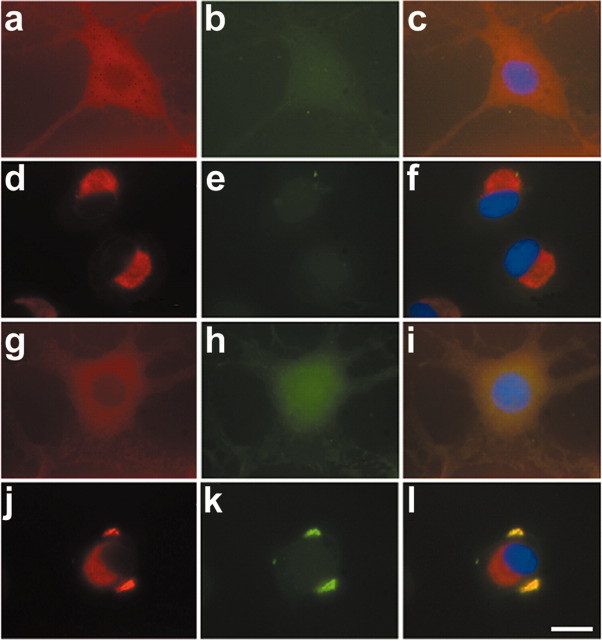
Figure 9.
Accumulation of cytoplasmic inclusions by proteasome inhibition after OA treatment. OLN-t40 cells were subjected to 0.5 μm MG-132 for 24 hr (d-f), to 20 nm OA for 24 hr (g-i), or to 20 nm OA for 6 hr followed by 0.5 μm MG-132 for 18 hr in the absence of OA (j-l). Indirect immunofluorescence was performed using MAb anti-αB-crystallin (red) followed by thioflavin-S staining (green). For nuclear staining (blue), DAPI was included in the mounting medium. a-c, Untreated control. a, d, g, j,αB-crystallin (red). b, e, h, k, Thioflavin-S (green). c, f, i, l, Overlay with DAPI. Scale bar, 20 μm.

