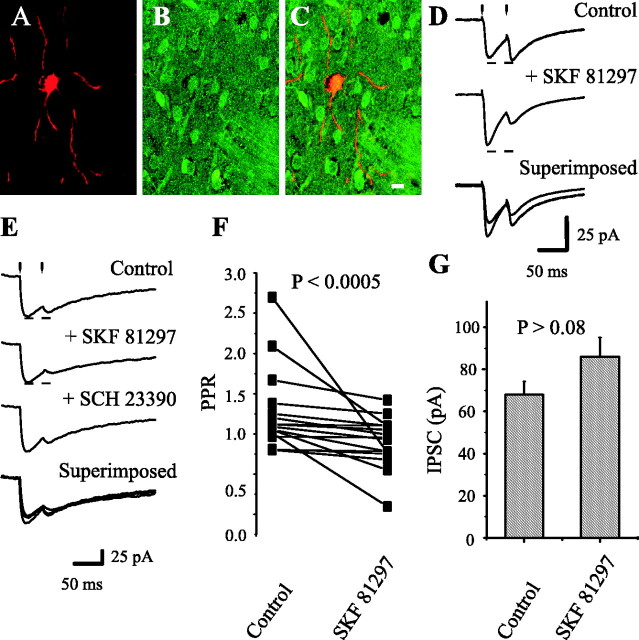
Figure 6.
Dopaminergic modulation of medium spiny axon collaterals. Activation of D1 receptors. Stimulating and recording electrodes as in Figure 1C. A, Neostriatal neuron filled with biocytin. B, Same preparation showing neurons immunoreactive for ENK. C, Superimposition of A and B with confocal microscopy; recorded neuron was ENK-positive. D, Top to bottom, First control synaptic current (in CNQX + AP5) was enhanced (12 of 16) by 100 nm SKF 21897, whereas PPR was decreased (14 of 16). Bottom trace shows superimposition of top and middle traces. E, SCH 23390 (100 nm) reverses the action of SKF 81297 in another cell. F, Paired line graph illustrates PPR reductions in a sample of spiny neurons (p < 0.0005; n = 16). G, Using the same stimulus strength, the mean of IPSC amplitudes (first response of the pair) before and during SKF 81297.

