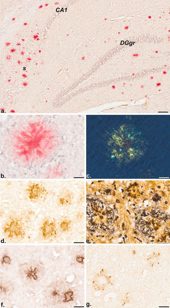
Figure 3.

Distribution of Aβ deposits revealed by histochemistry (a-c; Congo red with hemalum counterstain) and immunohistochemistry (d; BAP-2 antibody). Note the discrete congophilic staining of Aβ deposits in the hippocampal formation of a 17-month-old mouse (bright-field image a). The birefringent nature of a congophilic deposit (b) is demonstrated by polarization microscopy (c). Immunoreactive (BAP-2 positive) deposits in the neocortex of a 9-month-old mouse are illustrated in d. Plaque-associated dystrophic neurites are visualized by a modified cupric-silver staining procedure of de Olmos (Carlsen and De Olmos, 1981) (e). Immunoreactivities in f and g reveal the co-distribution of apoE and phospo-tau, respectively, with plaques. CA1, Ammon's horn layer 1; DGgr, dentate gyrus granule cell layer;. s, subiculum. Scale bars: a, 100 μm; b-g, 25 μm.
