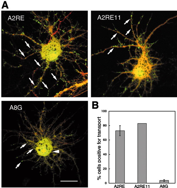
Figure 4.
Transport of A2RE-containing RNAs visualized in live neurons. Hippocampal neurons were microinjected with fluorescently labeled RNA (green) and Texas Red-labeled 10 kDa dextran (red), and after 30 min their distribution was determined by dual-channel confocal microscopy. A, Images of neurons microinjected with RNA. Exogenous RNAs containing A2RE or A2RE11 were observed to assemble into granules and move along the processes (arrows), whereas the A8G RNA did not assemble into granules but was distributed diffusely in the soma and proximal neurites (arrowhead) with only an occasional granule visible in the processes (arrows). Scale bar, 10 μm. B, Percentage of transport-positive cells for each of the microinjected RNAs in A. Error bars represent the SDs. For A2RE and A8G RNA, >20 cells were imaged for analysis. No error bar is given for A2RE11 because the number of cells imaged was small.

