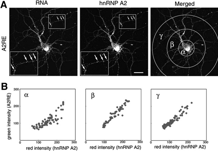
Figure 7.
hnRNP A2 colocalization with injected A2RE-containing RNA in granules. Differentiated hippocampal neurons were microinjected with fluorescently labeled RNA. Mouse antibody to hnRNP A2 and TRITC-labeled secondary antibody were used to locate this protein after fixing the cells. The subcellular distributions of the injected RNA and hnRNP A2 were visualized and analyzed by dual-channel confocal microscopy. A, Confocal microscopy images of neurons microinjected with RNA containing A2RE. Insets, At left, higher magnifications of cell areas marked by squares at right to demonstrate the colocalization of hnRNP A2 and A2RE-containing RNAs (arrows). Scale bar, 15 μm. The merged images are shown at right, with the areas selected for fluorescence intensity measurements of granules indicated. B, Analysis of relative RNA- and hnRNP A2-associated fluorescence in individual granules. α, β, and γ indicate the regions in the merged images from A used for each of the analyses.

