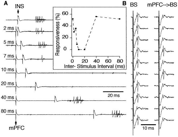
Figure 3.
Inhibition of a CeM output neuron by prestimulation of mPFC in the rat. A, CeM output neuron was synaptically driven by stimulation of the insular cortex (INS). Prestimulation of mPFC reduced the responsiveness of this CeM neuron to insular cortex stimulation (each trace shows 10 overlapping sweeps). Interval between mPFC and insula stimuli is indicated on the left. Inhibition lasted for up to 40 msec in this cell (inset). In the inset, responsiveness was calculated from 20 trials at each ISI. B, Inhibition of antidromic responses of a CeM output neuron. Each trace is the response to a single stimulus. The left column shows responses to brainstem (BS) stimuli applied in isolation. Prestimulation of mPFC (right column) reduced the probability of antidromic invasion of this CeM neuron from 80 to 20% (calculated from 20 trials). The ISI was 20 msec in this case.

