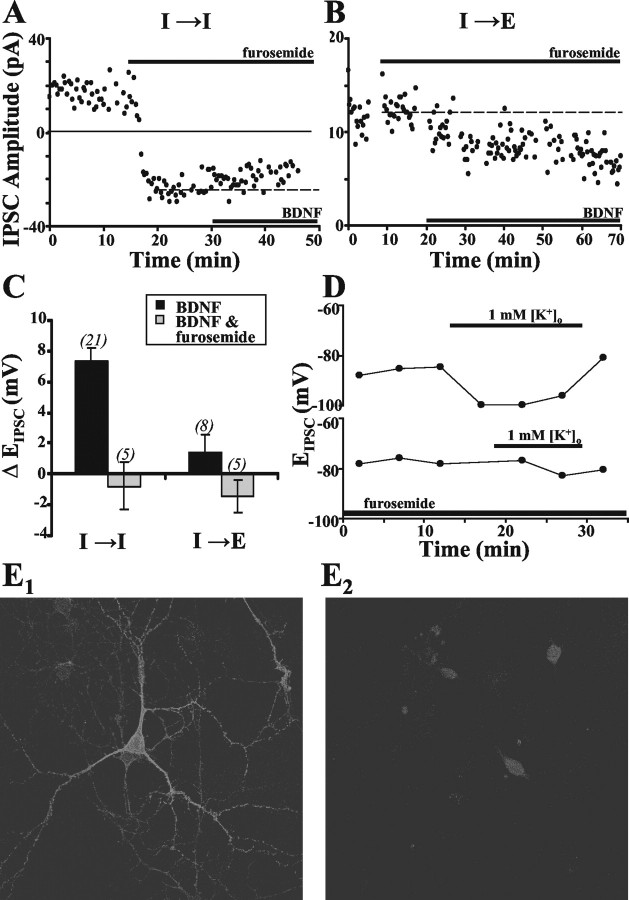
Figure 5.
Effect of furosemide on BDNF-induced modulation of EIPSC. A, B, Example recording of IPSCs (amphotericin B) at I→I and I→E synapses, showing the effects of furosemide (100 μm) and subsequent BDNF treatment. The dashed line shows the mean IPSC amplitude during the last 10 min before adding BDNF. C, Summary plots comparing BDNF-induced changes in EIPSC in the absence (black) or presence (gray) of furosemide at I→I and I→E synapses. Data obtained using both amphotericin B and gramicidin D showed no difference and were thus pooled. D, Two example recordings (gramicidin D) showing changes in EIPSC attributable to [K+]o-induced activation of KCC2 cotransport in the absence (top) and presence (bottom) of furosemide. Normal [K+]o (3 mm) was replaced with 1 mm [K+]o at the times indicated. E, Images of immunostained cells in hippocampal cultures. E1, Specific staining for KCC2 in these neurons using a KCC2 antibody as the primary antibody with cell-to-cell variation in KCC2 staining intensity. E2, Control staining in parallel cultures showing the level of nonspecific staining with the secondary antibody alone. For the control image, fluorescence intensity gain was increased more than twofold compared with that for specific KCC2 image. Nonspecific staining was restricted to the soma, showing no variation among neurons.

