
Figure 1.
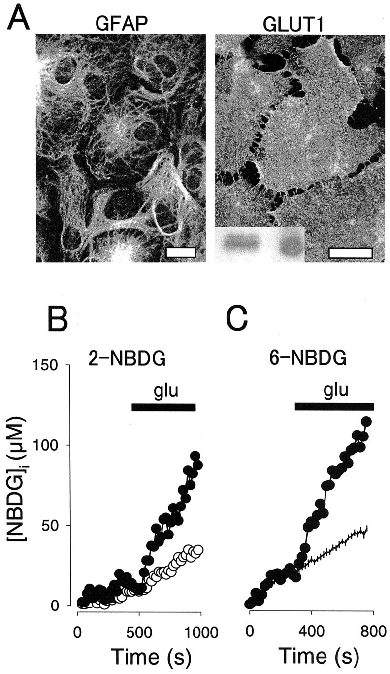
Glutamate stimulates the uptake of fluorescent hexoses by single hippocampal astrocytes. A, Cells were stained with antibodies against GFAP (left) or GLUT1 (right) and then imaged by confocal microscopy, as described in Materials and Methods. Scale bars, 20 μm. The inset shows GLUT1 immunodetected in 50 μg of total hippocampal proteins (left) and 50 ng of human erythrocyte membranes (right). B, Intracellular 2-NBDG was measured in two neighboring astrocytes during continuous exposure to 300 μm extracellular sugar. At the time indicated, 0.5 mm glutamate (glu) was added to the cells, resulting in different degrees of stimulation. C, Intracellular 6-NBDG was measured in a single astrocyte during continuous exposure to 300 μm extracellular sugar (filled symbols). At the time indicated, 0.5 mm glutamate was added. Data from 20 nonstimulated control cells are shown for comparison (mean ± SEM).