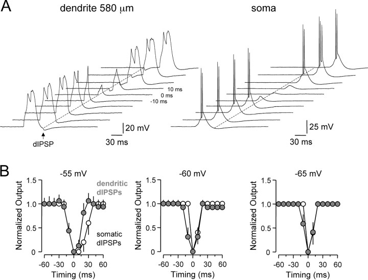
Figure 9.
Voltage-dependent inhibition of dendritic spike initiation. A, Tiled records of dendritic spike firing evoked by short dendritic current steps (from -55 mV), offset in time by 10 msec, recorded simultaneously at dendritic (left) and somatic (right) sites. dIPSPs generated at the dendritic site sculpt and suppress dendritic spike generation and consequent axonal action potential initiation. Timing = 0 represents coincidence of the dendritic current step and dIPSC onset. B, Comparison of the time window for inhibition of normalized axonal spike output by dIPSPs. Open symbols show the time window for suppression of axonal spike generation by somatically generated dIPSPs. The gray symbols show the time window for suppression of axonal action potential firing, generated as a consequence of the forward propagation of dendritic spikes, by dendritic dIPSPs. Data are shown for three membrane potentials (-55, -60, and -65 mV). Points represent normalized mean ± SEM. Note the time window for inhibition of neuronal output is significantly longer for somatically generated dIPSPs only at the most depolarized membrane potential tested.

