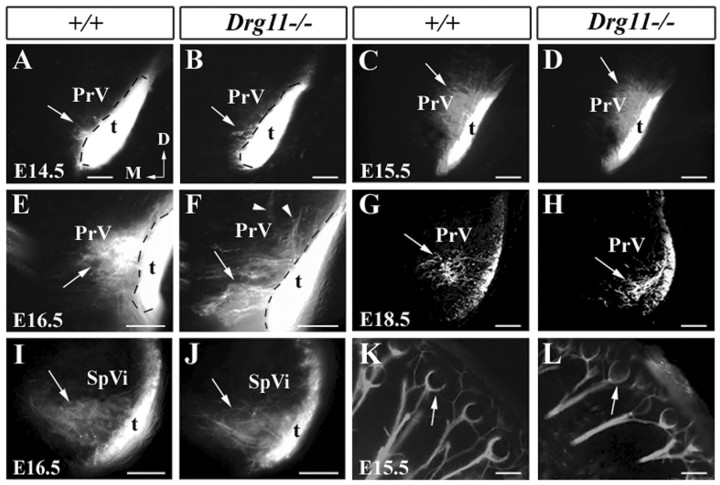
Figure 4.
Central and peripheral projections of TG afferents in wild-type and Drg11 mutant embryos as revealed by DiI staining. A,B, At E14.5, both wild-type (A) and mutant (B) TG afferents enter the PrV (arrows). Note that DiI crystals were applied to the b2 vibrissal follicle. C,D, At E15.5, more DiI-labeled axons are observed within the wild-type (C) and mutant (D) PrV. Most afferents have a dorsomedial trajectory (C,D, arrows). No major abnormality is noted in the mutant (D). Note that DiI crystals were applied to the c2 and d2 vibrissal follicles. E,F, At E16.5, DiI-labeled wild-type afferents further reach the inner region of the PrV with a dorsomedial trajectory (E, arrow). The mutant afferents display ventromedial trajectories (F, arrow). A few afferent fibers grow aberrantly in a dorsolateral direction (F, arrowheads). Note that DiI crystals were applied in the b2 and b3 vibrissal follicles. G,H, At E18.5, TG afferents become clustered (G, arrow) in the inner region of the PrV in wild-type mice (G, arrow). By contrast, mutant afferent clusters are located in more ventrolateral regions (H, arrow). Note that DiI crystals were placed in the c2 vibrissal follicle. These images were obtained through the use of a laser confocal microscope. I,J, No difference in the projection patterns of TG afferents is found between the SpVi of wild-type (I, arrow) and mutant (J, arrow). Note that DiI crystals were placed in the b2 and b3 follicles. K,L, DiI tracing of TG afferents to the vibrissal follicles of E15.5 wild-type (K, arrow) and mutant (L, arrow). Note that DiI-labeled fibers surround the base of the follicles and exhibit a circumferential profile (K,L, arrows). Scale bars, 100 μm.

