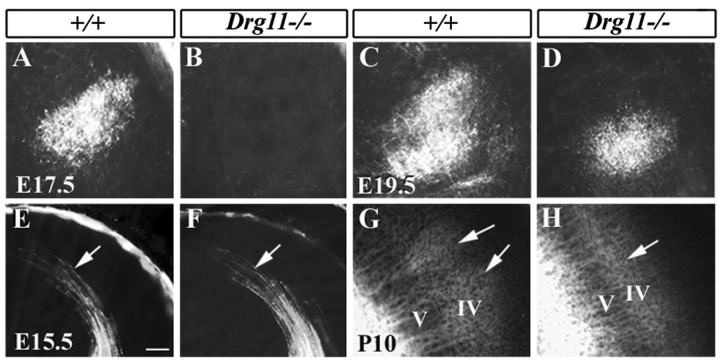
Figure 6.
Projections of PrV efferents and TCAs revealed by DiI tracing. A-D, DiI tracing of PrV projections in the VPm. A,B, Whereas PrV efferents are present in the E17.5 wild-type VPm (A), no DiI-labeled axons are present in the mutant VPm (B). C,D, At E19.5, DiI-labeled PrV efferents are found in both the wild-type (C) and mutant (D) VPm, but the extent of DiI labeling in the mutant is much reduced (D). E-H, DiI tracing of TCAs in the S1 cortex. E,F, At E15.5, TCAs of wild-type and mutants labeled by DiI are found in the S1 cortex. Arrows point to the growth cones of TCAs that arrive at S1 cortex. No major difference is found in the projection patterns. G,H, The projection of TCAs in layer IV of the cortex at P10. In wild-type mice (G), DiI-labeled axons aggregate to form individual patches corresponding to single barrels (G, arrows), but in the mutant (H), DiI-labeled axons are distributed evenly. Scale bar, 100 μm.

