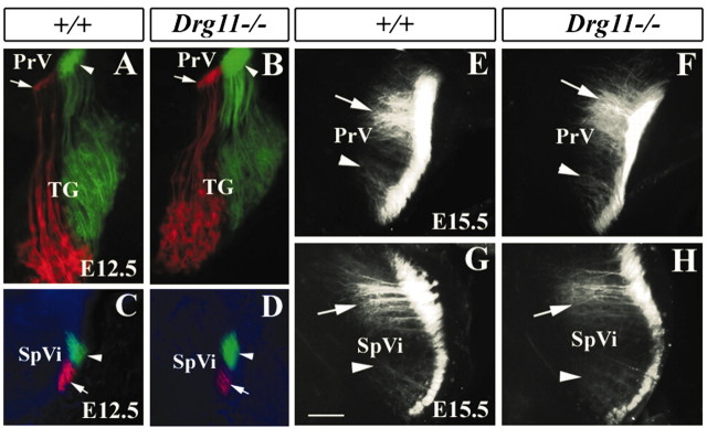
Figure 7.
Topographic organization of the TG projections revealed by DiI and DiA double labeling. A-D, DiI and DiA were applied in the dorsal and ventral vibrissae follicles of wild-type (A,C) and mutant (B,D) embryos, respectively, at E12.5. DiI-labeled (red; arrows) and DiA-labeled (green; arrowheads) axons are segregated in the TG (A,B), PrV (A,B), and SpVi (C,D). No major difference between the wild type and mutant is noted. E,H, DiI crystals were applied to distinct vibrissae follicles of wild-type (E,G) and mutant (F,H) embryos, respectively, at E15.5. Two well separated foci of DiI-labeled afferents mark distinct clusters of TG afferents in the PrV (E,F, arrowheads) and SpVi (G,H, arrowheads). Note the equivalent locations and densities of labeled afferents in the wild type and mutants. Scale bar, 100 μm.

