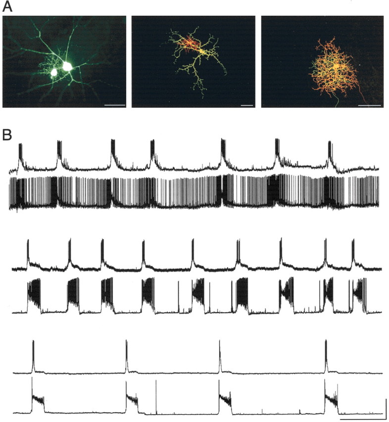
Figure 6.

Synchronous firing of neighboring morphologically identified ganglion cells. A, Three examples of filled cell pairs. The first photomicrograph shows an α- and a β-cell (age, P21) filled with Lucifer yellow, as was done in early experiments. In subsequent experiments, two distinct dyes were used [Alexa fluor 568 (red) and 488 (green)] to identify individual cells. Two examples are shown [β-γ, middle age (P16); γ-γ, right (P22)]. Scale bars, 50 μm. B, Recordings from neighboring cell pairs obtained after morphological and physiological differentiation demonstrate that neighboring ganglion cells of all morphological classes burst in a coordinated manner regardless of cell type. The first pair of traces includes a β-cell (top; Vm = -68 mV) and an α-cell (bottom; Vm = -66 mV) from a P21 animal. The second pair includes a γ-cell (top; Vm = -63 mV) and a β-cell (bottom; Vm = -72 mV) from a P19 animal. The spontaneous activity of two γ-cells from a P18 animal is shown in the last pair of traces (Vm for both = -59 mV). Calibration: x, 30 sec; y, 40 mV.
