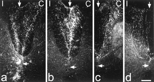
Figure 6.
BD labeling in the dorsal columns. Sections of the spinal cord close to the injection sites are shown for the four experiments in which BD was injected into the dorsal columns. BD has been detected with streptavidin-rhodamine, and each image shows a single optical section scanned to reveal this with the confocal microscope. In each case the left side is ipsilateral (I) and the right side is contralateral (C) to the left sciatic nerve, which had been transected between 8 and 11 weeks earlier. The top vertical arrow indicates the midline, and the bottom horizontal arrow indicates the position of the central canal. In a and b successful bilateral labeling was achieved. In a one of the injection sites had passed ventrally through lamina X and into the region of the ventral white commissure. c and d are from experiments in which only one injection was successful. In c, this was on the right (contralateral) side and in d it was on the left (ipsilateral) side. Scale bar, 200 μm.

