
Figure 5.
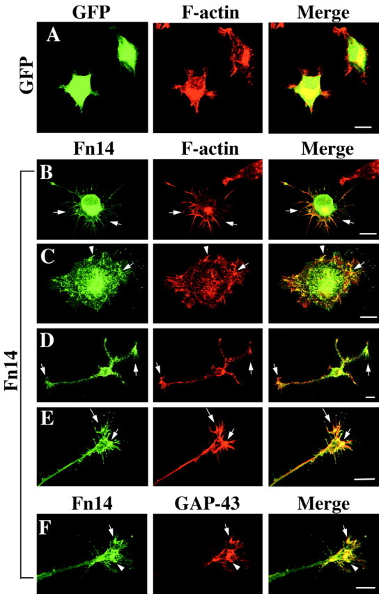
Fn14 overexpression in PC12 cells enhances filopodial and growth cone formation. A-E, PC12 cells were cultured for 24 hr after infection with HSV-GFP or HSV-Fn14wild-GFP and examined for GFP (green) and F-actin (red, rhodamine-phalloidin). Although HSV-GFP has no effect on cell morphology (A), overexpression of Fn14 induces a number of long filopodia extending from straight cell borders and from angular cell borders (B). Spreading and lamelipodial formation is also observed in Fn14-expressing PC12 cells (C). Filopodia and lamelipodia are seen at the distal ends of formed growth cones (D, E). Fn14 and F-actin are well colocalized near the cell perimeter and in filopida and lamelipodia (arrows). F, After HSV-Fn14wild-GFP infection, GAP-43 (red) is expressed abundantly in filopodia and lamelipodia and colocalized with Fn14 (arrows). Scale bars, 10 μm.