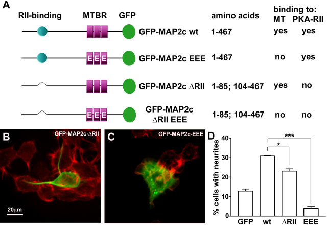
Figure 7.
Functional significance of MAP2c-interacting proteins during neurite initiation. Neuro-2a cells were transfected to express variants of GFP-MAP2c (green), fixed, and stained for F-actin (red). A, Domain diagram depicting MAP2c mutants used in this study. B, Cells transfected with GFP-MAP2c-ΔRII were able to induce neurites; however, the efficiency was significantly reduced. C, Pseudophosphorylated MAP2c (GFP-MAP2c-EEE), which is strongly impaired in microtubule binding, failed to induce neurites. D, Quantification of neurite formation. Data represent three independent experiments, each with >100 cells evaluated per treatment condition (*p < 0.05; ***p < 0.001, one-way ANOVA). MT, Microtubules; MTBR, microtubule binding repeats; wt, wild type.

