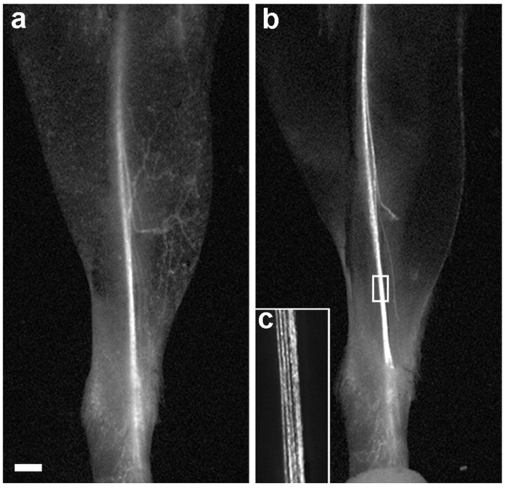
Figure 1.
Transcutaneous imaging of the saphenous nerve. a, The leg of a YFP-H transgenic mouse imaged with a fluorescence dissecting microscope after depilation. The saphenous nerve and some of its cutaneous branches are visible. b, After the image in a was captured, the mouse was killed, the skin was removed, and the leg was imaged again. The boxed area in b is shown at higher magnification in c. Scale bar, 1 mm.

