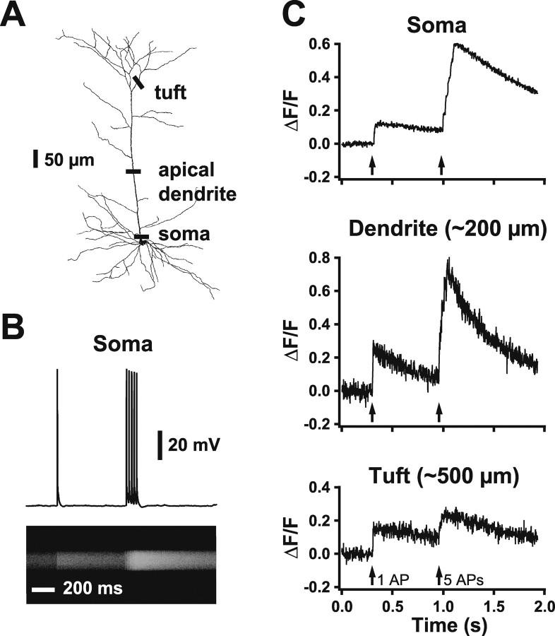
Figure 8.
Backpropagating somatic APs cause calcium transients in the apical dendrites of prefrontal neurons. A, A reconstructed prefrontal layer 5 pyramidal neuron showing somatic, dendritic, and tuft locations of line scans used to detect calcium signals in response to somatic APs. B, In a cell filled with Oregon Green BAPTA-1 (200 μm), somatic current injection produces APs (top) that result in a calcium-dependent change in fluorescence imaged using a line scan at the intersection of the soma and apical dendrite (bottom). C, Plot of ΔF/F in response to a similar spike train imaged at three locations: the intersection of the soma and apical dendrite (top), in the apical dendrite (middle; ∼200 μm from the soma), and in a proximal branch of the apical tuft (bottom; ∼500 μm from the soma). Data are from three different neurons.

