
Figure 9.
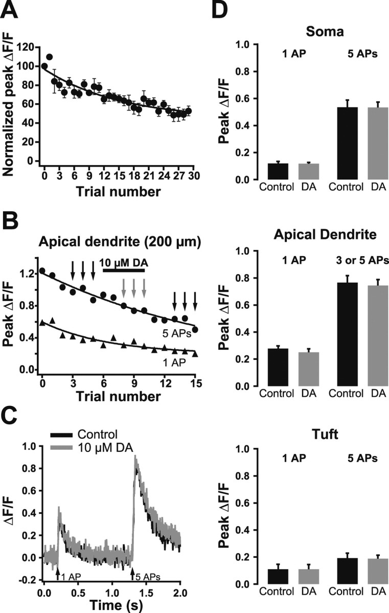
Dopamine does not modulate calcium signaling in the apical dendrite. A, Plot of normalized average peak ΔF/F (±SEM; n = 3) in response to somatic APs imaged 200 μm from the soma. Note the time-dependent decrease in ΔF/F. B, Plot of peak ΔF/F during single (triangles) and trains of five APs (circles) during dopamine (DA) application at the indicated time (bar). C, Comparison of averaged ΔF/F traces in control conditions (black; before and after dopamine) and during dopamine application (gray). Traces shown are averages of trials at the times indicated by the black and gray arrows in B. D, Summary graphs showing a lack of effect of dopamine on dendritic calcium transients evoked by single APs or trains of five APs at the soma (top), 200 μm into the apical dendrite (middle), and in the proximal apical tuft (bottom).