Figure 2.
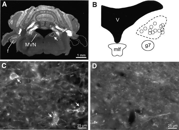
Distribution of Purkinje cell terminal outlines after unilateral flocculectomy. A, GFP signal in a coronal section of an adult mouse in which the right flocculus (f) and paraflocculus were surgically ablated. The dashed line on the right outlines the portion of the cerebellum that was ablated. The region of the MVN encircled on the right side (arrow) corresponds with the dashed region in B. B, Schematic of the MVN. The dashed line encircles the region in which terminal outlines were missing on the side ipsilateral to the lesion, as exemplified in D. The circles indicate the approximate locations of intracellularly recorded FTNs described in this study. C, High-magnification view of the center of the region outlined in B, taken on the side contralateral to the floccular lesion. Three FTNs are evident (arrows). D, The same location as in C, but on the side ipsilateral to the lesion, is devoid of terminal outlines. Scale bars: A, 1 mm; C, D, 25 μm.

