Figure 1.
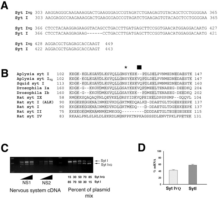
Cloning of a novel spliced isoform of Syt I. A, Nucleotide sequence of two clones amplified from a nervous system library showing the insertion of six amino acids. Nucleotide numbering is from residues 303–449. B, Alignment of juxtamembrane domain from a number of species highlighting the conservation of this region in Syt Is and the conservation of the spliced forms. Drosophila Ib (Dros Ib) (from EST clones; accession numbers 15484159, 15504610, and 15505802) and Syt IALK (Perin et al., 1990). Syt IX has also been called Syt V in other publications. The black bar represents the site of alternative splicing, and the star represents the site of PKC phosphorylation. C, RT-PCR of Syt I and Syt IVQ demonstrates approximately equal amounts of both splice forms. The insertion of the VQ generates an RsaI site. We used PCR primers flanking the insert for RT-PCR from the Aplysia nervous system. The amplified product was then cut with RsaI to determine the proportion of RNAs with the insert. Different amounts of nervous system template were used to ensure that PCR amplification was in the linear range. Results are shown for two different animals (NS1 and NS2). To generate a standard curve, mixes of plasmids containing different proportions of Syt IVQ and Syt I were used as the template for PCR. D, The proportion of the two RNAs was calculated based on the standard curves. Values are mean ± SEM for four independent RT-PCRs from four individual animals.

