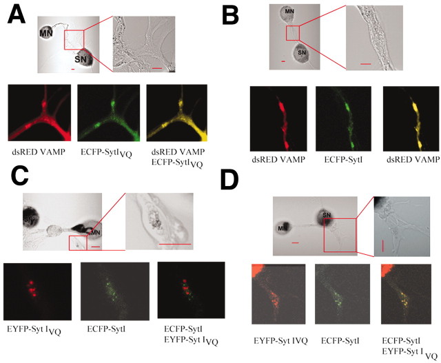
Figure 2.
Localization of Syt I and Syt IVQ in sensory neurons. Plasmids encoding DsRed-labeled Aplysia VAMP and ECFP-labeled Syt I (A) or DsRed-labeld Aplysia VAMP and ECFP-labeled Syt IVQ (B) were injected into sensory neurons. Expressing neurons were then paired with motor neurons and visualized 3–5 d later. For five cells expressing Syt I and four cells expressing Syt IVQ, all DsRed VAMP clusters were completely overlapped with ECFP-Syt I (43 clusters) or ECFP-Syt IVQ (14 clusters). C, Plasmids encoding ECFP-labeled Syt I and EYFP-labeled Syt IVQ or ECFP-labeled Syt IVQ and EYFP-labeled Syt I were injected into sensory neurons. Expressing neurons were then paired with motor neurons and visualized 3–5 d later. D, In the majority of images, EYFP Syt IVQ and ECFP Syt I did completely overlap. Scale bars, 20 μm.

