
Figure 3.
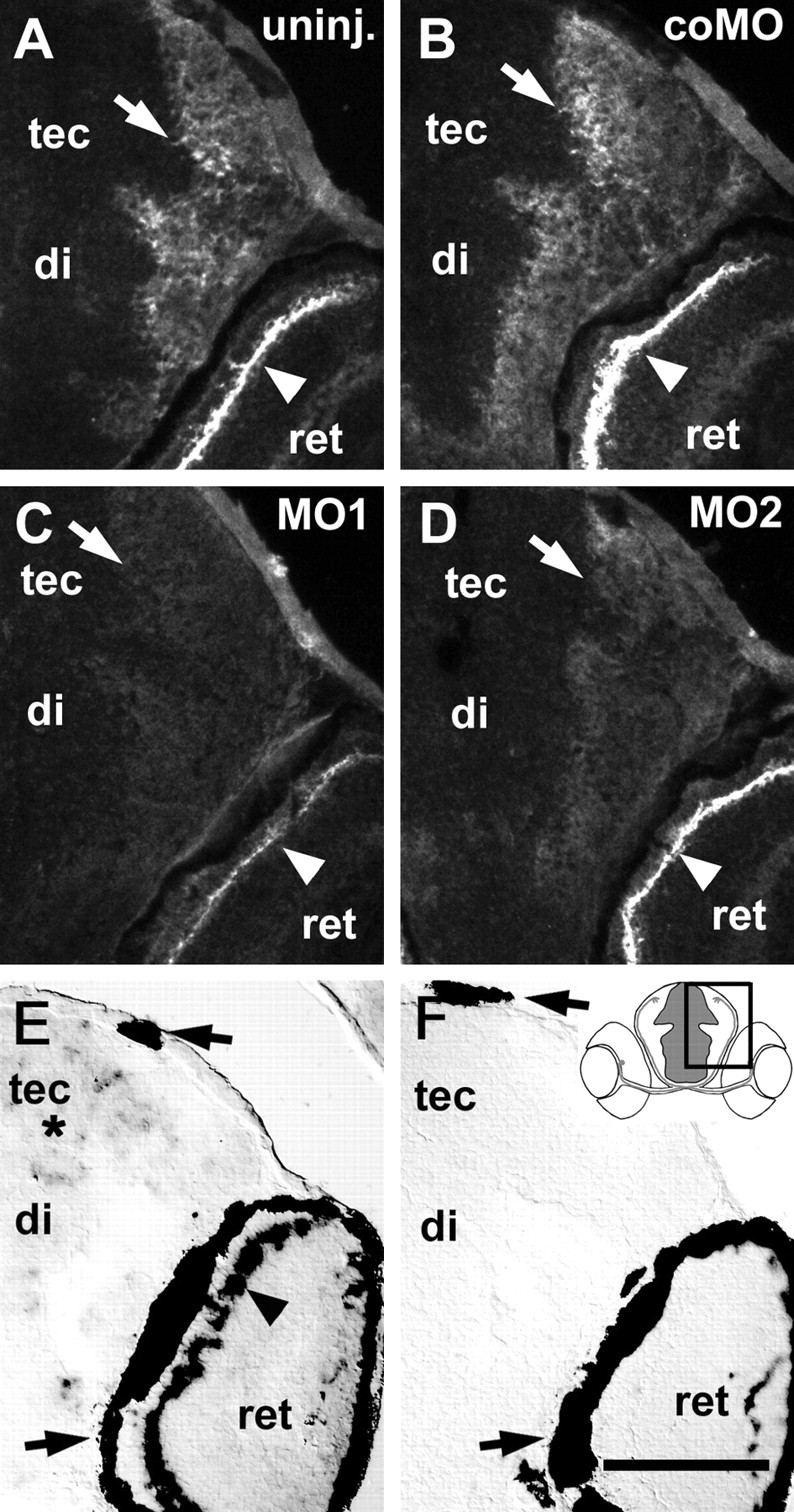
Effect of morpholinos on tenascin-R immunoreactivity and tenascin-R mRNA expression in 3-d-old larvae. Cross-sections including the diencephalon (di), tectum (tec), and part of the retina (ret) correspond to the boxed area in the inset in F. A–D, Arrows depict deep tectal fiber layers, and arrowheads indicate the outer plexiform layer of the retina. In uninjected (A, uninj.) and control morpholino-injected (B, coMO) larvae, tenascin-R immunoreactivity is most intense in the outer plexiform layer of the retina and high in deep fiber layers of the tectum and diencephalon. Injection of morpholino1 (C, MO1) more strongly reduces immunoreactivity than morpholino2 (D, MO2). E, F, Tenascin-R mRNA is detected by in situ hybridization in scattered cells throughout the periventricular cell layer of the brain (asterisk) and in a row of cells in the retina (arrowhead in E) but not after incubation with a sense RNA probe (F). Arrows in E and F indicate the retinal pigment epithelium and pigment cells in the epidermis, which always appear black. Scale bar (in F): A–F, 100 μm.