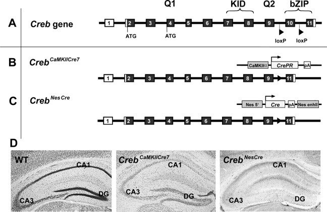
Figure 1.
Schematic representation of the targeting strategy used for the generation of mouse strains with a progressive reduction of CREB in the brain. A, Genomic structure of the Creb gene. Coding exons are indicated by filled boxes; noncoding exons are indicated by open boxes. 5′- and 3′-flanking sequences and introns are given as lines. Q1, KID, Q2, bZIP, Domains of the CREB protein; Q, glutamine-rich transactivation domain; b, basic region; ZIP, leucine zipper dimerization domain; ATG, start condon. B, CrebCamKCre7, LoxP-flanked Creb exon 10 was excised by a constitutively active Cre-recombinase fused with the C-terminal-truncated ligand binding domain of the progesterone receptor fusion (Kellendonk et al., 1996) expressed under the control of a CamKIIα promoter. Filled triangles indicate the loxP site remaining after excision of exon 10. pA, Polyadenylation signal. C, CrebNesCre, Exon 10 was deleted under the control of the brain-specific promoter nestin (Nes). enhII, Enhancer II. D, CREB immunostaining in the different Creb mutant and wild-type lines at 8–12 weeks of age. WT, Wild type; DG, dentate gyrus.

