Figure 3.

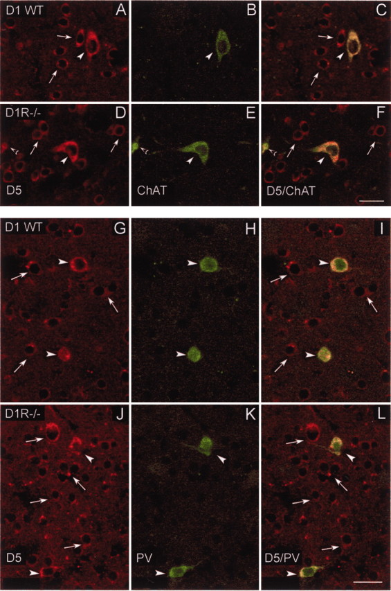
Confocal laser photomicrographs illustrating the colocalization of D5 receptors with choline acetyltransferase (ChAT) and parvalbumin (PV) in striatal interneurons of WT and DA D1R-/- mice. A, D, Striatal neurons expressing DA D5 receptors in WT (A) and in D1R-/- mice (D); B, E, striatal interneurons containing ChAT in WT (B) and in D1R-/- mice (E). C and F show paired images illustrating double-labeled cells with D5/ChAT in WT (C) and D1R-/- mice (F). Note in C and F that ChAT-positive neurons express D5 receptors in WT and D1R-/- mice. Open arrowheads in D–F indicate a cholinergic partial cell that also expresses D5 receptors. G, J, Striatal neurons expressing DA D5 receptors in WT (G) and in D1R-/- mice (J); H, K, PV-containing interneurons located in the striatum of WT (H) and D1R-/- mice (K). Paired images in I and L show double-labeled cells with D5/PV in WT (I) and D1R-/- mice (L). White arrows indicate single-labeled cells for D5 receptors, and arrowheads indicate double-labeled cells in the corresponding images. Note in I and L that PV-containing neurons also express D5 receptors in WT and in D1R-/- mice. Scale bars, 25 μm.
