
Figure 1.
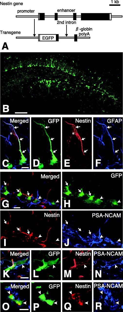
GFP-expressing cells in the dentate gyrus of adult nestin promoter-GFP mice. A, Structure of nestin promoter-GFP transgene. The nestin promoter-GFP transgenic mouse expressed GFP under the regulatory element of nestin promoter and the second intron. B, The dentate gyrus of the nestin promoter-GFP mouse observed with the aid of a confocal microscope. C-F, The nestin immunostaining to the GFP-expressing cells with the process bearing GFAP. C, Merged image. D, GFP fluorescence; E, immunostaining for nestin; F, immunostaining for GFAP. The cell expressing nestin also expressed GFAP (arrows) and often comes in contact with blood vessels, which were also positively stained with anti-nestin (E; asterisk) as described previously (Palmer et al., 2000), but this blood vessel did not express GFP signal. G-R, The three sets of the confocal image (G-J, K-N, and O-R) demonstrating the nestin immunostaining to the GFP-expressing cells bearing PSA-NCAM proteins at their cell surface (arrowheads). The merged image is shown in G, K, O; GFP fluorescence is shown in H, L, P; the immunostaining for nestin protein is shown in I, M, Q; the immunostaining for PSA-NCAM is shown in J, N, R. Note that the cell surface areas of PSA-NCAM-bearing GFP-positive cells were positively stained with this anti-nestin antibody. Scale bars: B, 100 μm; C-R, 10 μm.