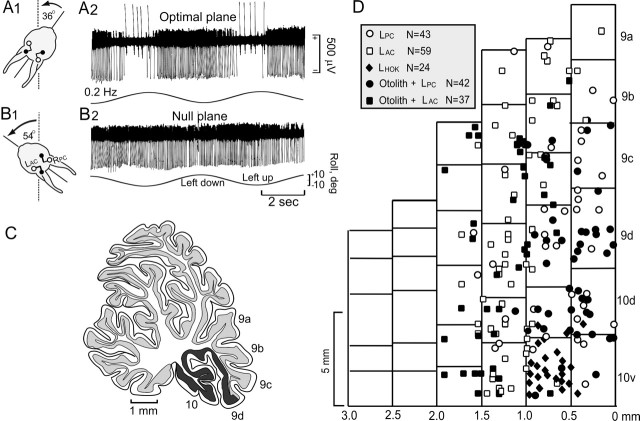
Figure 2.
Topographic distribution of CFR optimal planes within uvula-nodulus. Sinusoidal vestibular stimulation was used to classify optimal response planes of CFRs. A, B, Figurines illustrate optimal (A1) and null (B1) CFR response planes of a Purkinje cell recorded in the left uvula. Stimulation in the optimal plane (A2) evoked increased CFRs and decreased SSs when the rabbit was rotated onto its left side. When stimulated in the null plane (B2), neither CFRs nor SSs were modulated. C, Sagittal view of the rabbit cerebellum. The shaded area (folia 9d and 10) indicates the region of the uvula-nodulus receiving vestibular primary afferent projection. D, Folia 9-10 are represented as a two-dimensional topographic sheet. Optimal planes for CFRs recorded from 205 Purkinje cells are represented on this surface. Cells with optimal planes corresponding to stimulation in the plane of the ipsilateral posterior semicircular canal (LPC) are illustrated as circles. Cells with optimal planes corresponding to stimulation in the plane of the ipsilateral anterior semicircular canal (LAC) are illustrated as squares. Open symbols indicate cells in which the optimal plane was determined only for sinusoidal stimulation. Filled symbols indicate cells that were tested for static sensitivity and were positive. Black diamonds indicate cells that responded only to horizontal optokinetic stimulation (LHOK) of the ipsilateral eye in the posterior-anterior direction. The more lateral aspects of folia 9-10 were unexplored.

