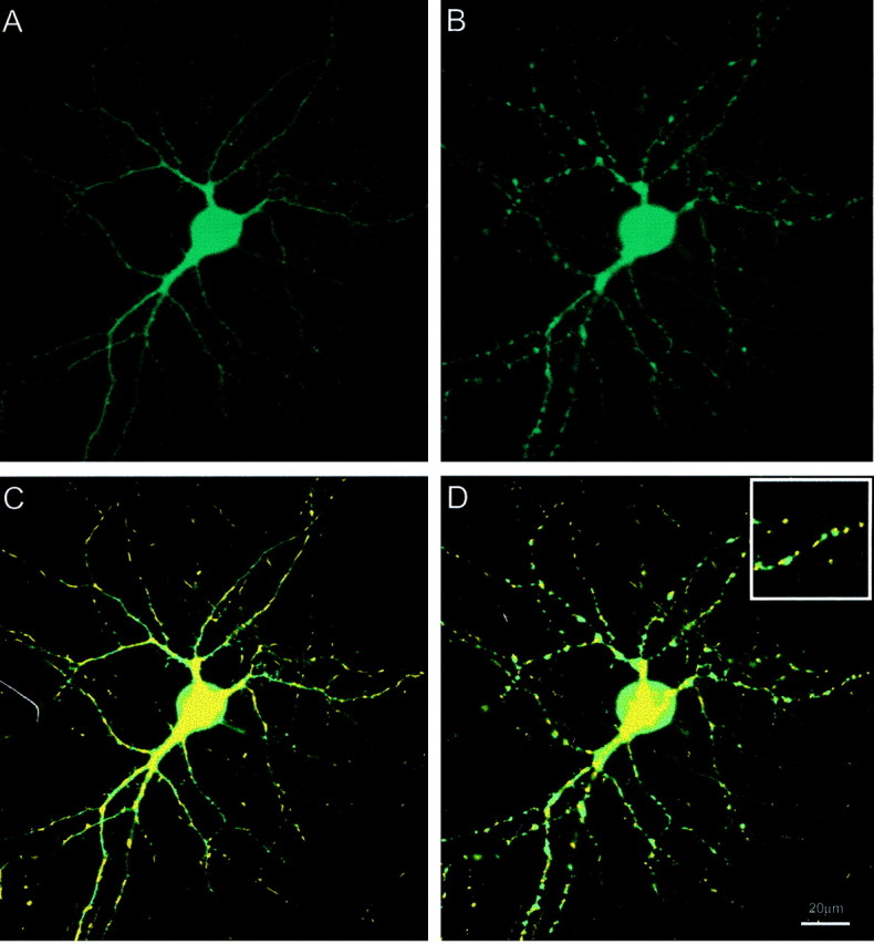
Figure 6.

Comparison of mitochondrial and dendrite remodeling. A, Single cortical neuron transfected with cytosolic eCFP. B, Effect of perfusion on dendrite morphology after 5 min perfusion with 30 μm glutamate/1 μm glycine. C, Overlay of fluorescence images of mt-eYFP and cytosolic ECFP before glutamate exposure. D, Overlay image after superfusion with 30 μm glutamate/1 μm glycine. Inset, Magnified region. These data are representative of four additional fields.
