
Figure 4.
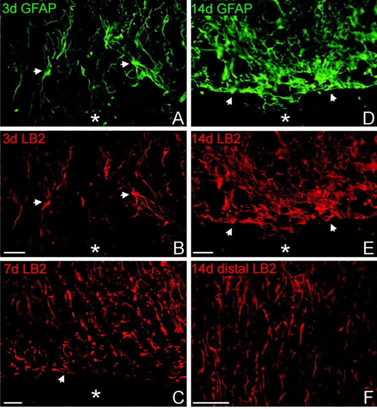
Reactive astrocytes at the lesion interface express ephrin-B2. Horizontal sections through lesioned tissue were double stained for GFAP (A, D; green) and ephrin-B2 (LB2; B, C, E, F; red) to demonstrate colocalization within astrocytes. Rostral is oriented toward the top of the page, and the lesion cavity is marked with an asterisk in each panel. The injured tissue immediately rostral to the lesion is depicted in A-E. A, B, At 3 d after injury, ephrin-B2-positive astrocytes were loosely distributed around the lesion area. A few hypertrophic astrocytes double stained for GFAP (arrows) had increased ephrin-B2 immunoreactivity. Astrocytic processes were disorganized and oriented radially toward the lesion but not parallel along the lesion surface. C, By 7 d after injury, many ephrin-B2-positive astrocytes had reoriented their processes parallel to the lesion interface (arrow). D, E, At 14 d after injury, astrocytes at the glial scar formed a dense cellular network with slender processes oriented parallel to the lesion surface. All astrocytes and astrocytic processes along the lesion surface possessed high levels of ephrin-B2 immunoreactivity compared with astrocytes in white matter several millimeters distal to the lesion site in the same tissue section (F). Scale bars, 50 μm.