
Figure 5.
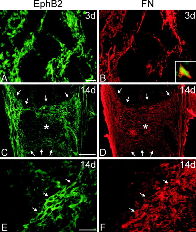
Immunohistochemistry for EphB2 after injury. Horizontal sections through the spinal cord lesion were double stained for EphB2 (A, C, E; green) and fibronectin (FN; B, D, F; red) to detect fibroblasts in the lesion cavity at 3 and 14 d after injury. In all panels, rostral is oriented toward the top of the page. A, B, At 3 d after transection, there was generally weak EphB2 staining in the necrotic regions of the cord surrounding the lesion cavity. However, a loose meshwork of cells positive for EphB2 (A) and fibronectin (B) infiltrated the lesion cavity along the lesion interface. These cells appeared to originate from the meninges. B, Inset, Higher magnification of a cell double stained for EphB2 and fibronectin. C-F, By 14 d after injury, the lesion cavity was filled with a dense meshwork of EphB2-positive cells. C, D, Low-magnification micrographs of horizontal tissue sections through the entire lesion site. Asterisks mark the lesion epicenter, in which there is reduced EphB2 staining (C), and arrows demarcate the lesion interface with the spinal cord. Although fibronectin-positive fibroblasts have filled the entire lesion cavity (D), those that are the most immunoreactive for EphB2 are concentrated along the spinal cord border and meninges. E, F, Higher magnification of EphB2-positive meningeal fibroblasts within the lesion. Arrows mark cells double stained for EphB2 and fibronectin at the lesion interface. Scale bars: A, B, E, F, 50 μm; C, D, 500 μm.