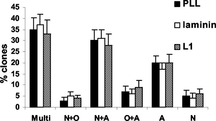
Figure 12.
Percentages of different clone types on PLL, laminin, or L1 substrate. After precursor cells were maintained on PLL, laminin, or L1 substrates for 15 d, the distribution of different clone types was scored for each substrate. No significant differences were found between different substrates. Values are means + SEM. Clone types were as follows: Multi, multipotential; N+O, neuronoligodendrocyte bipotential; N+A, neuron-astrocyte bipotential; O+A, oligodendrocyte-astrocyte bipotential; A, astrocytic monopotential; N, neuronal monopotential.

