
Figure 2.
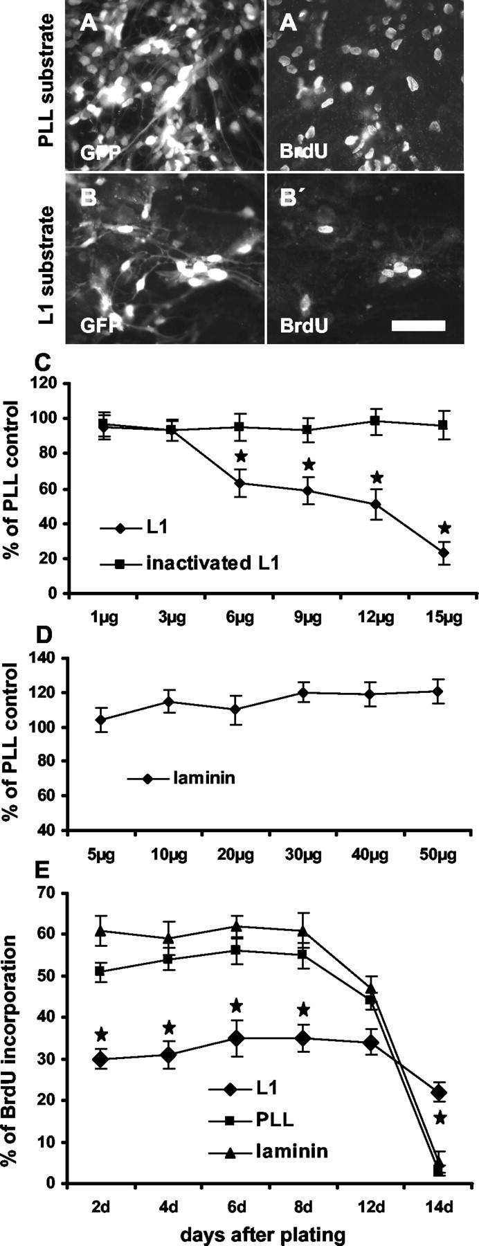
Substrate-coated L1 inhibits precursor cell proliferation. To measure the influence of different substrates on proliferation, dissociated precursor cells were plated on PLL, L1, or laminin substrates. Six days after plating, an 8 hr BrdU pulse was administered, and the percentage of BrdU+ cells was determined. Photomicrographs show more GFP+ precursor cells on PLL (A) than on L1 (B) substrate. BrdU+ cells are shown in corresponding figures of identical fields (A′, B′). C, Percentages of all cells having incorporated BrdU when grown on substrates consisting of different concentrations of L1 or heat-inactivated L1 are shown in relation to BrdU incorporation on PLL substrate, which was set to 100%. D, Percentage of BrdU-labeled precursor cells on laminin substrate at different concentrations is shown in relation to BrdU incorporation on PLL substrate, which was set to 100%. E, Time course of BrdU incorporation of precursor cells growing on the indicated substrates is shown. Decreasing amounts of BrdU+ cells were detected at time points that precursor cells reached 80-90% confluency. Scale bar, 50 μm. *p < 0.05 versus the corresponding time point on PLL substrate.