Figure 3.
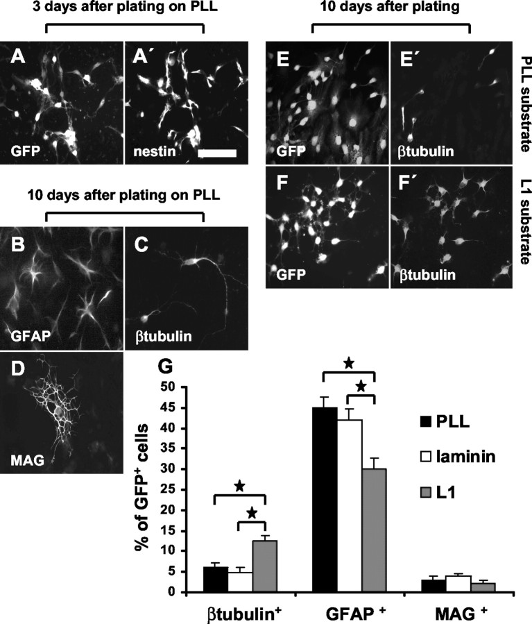
Substrate-coated L1 enhances neuronal yield after precursor cell differentiation. Three days after plating, in the presence of growth factors, most of the precursor cells remain nestin+, independent of the substrate. A, A′, GFP+ and nestin+ cells in the same microscopic field. At this time point, ∼90% of precursor cells express nestin. After differentiation by growth factor withdrawal, GFAP+ cells (B), β-tubulin+ cells (C), and MAG+ cells (D) were found. On L1 (6 μg/ml) substrate, the percentage of β-tubulin+ neurons is increased when compared with PLL substrate (E, E′, F, F′). On laminin (20 μg/ml) substrate, the percentage of β-tubulin+ neurons was similar when compared with PLL substrate (G). Results concerning differentiation of precursor cells are summarized in G. Scale bar, 50 μm. Values are means + SEM. *p < 0.05 versus the indicated bar.

